Eshealthtips.com – A physician may diagnose claw toe deformity through a physical exam and biomechanical examination. A podiatrist will check for stiffness, reduced range of motion, and pain associated with specific joint motions. X-rays may also be ordered to confirm a diagnosis. Treatment for claw toe can be conservative, but in severe cases, surgery may be necessary. Your podiatrist will determine whether treatment is necessary based on the type of deformity and your individual condition.
Causes of Claw Feet and Routine Treatment
There are many causes of claw toe, including genetics and foot type. People with flat or high-arched feet are more likely to develop claw toes. However, other disorders such as neuromuscular disease can exacerbate the condition. Wearing tight or pointed-toed shoes can also aggravate the deformity. Regardless of the cause, doctors recommend the use of appropriate footwear and regular care for calluses and corns to help minimize pain and discomfort.
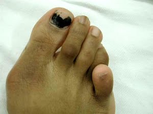
Claw toe deformities are caused by excessive pressure on the dorsum of the toe. This pressure causes the distal interphalangeal joint to permanently flex. Claw toe can affect any toe, except the great toe. Another form is known as mallet toe, which affects only the second toe. It is not uncommon for children to have this deformity, but is not completely curable.
Conservative treatment for claw toe deformity usually involves wearing a soft-toed shoe and doing toe exercises to strengthen the toes. In severe cases, hammertoe surgery is recommended. The initial treatment is often enough, but if left untreated, it can lead to permanent deformity. If left untreated, it can even result in nerve damage, weaken foot muscles, and painful calluses.
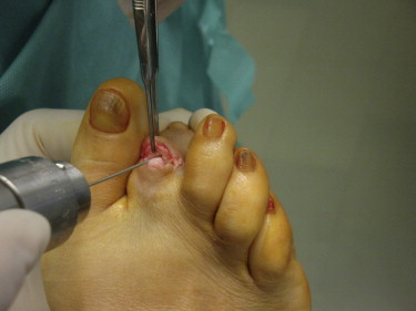
Surgical Treatment for Toe Claw Deformities
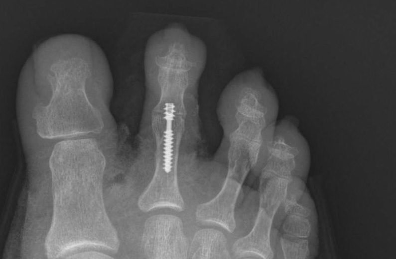
The Girdlestone-Taylor procedure is one surgical treatment for claw-toe deformities. The procedure involves dividing the FDL flexor to the extensor tendon. This technique has good success rates and reduces the risk of harness syndrome and excessive stiffness. While arthodesis has fewer complications than arthodesis, a cocked-up toe is still a cosmetic concern for many patients.
The root cause of claw toe deformity is a dysfunctional MTP joint. Repetitive loading of the joint over time causes the intrinsics to shorten, making it inefficient in producing a flexion moment. The resulting instability can result in pain and instability in the MTP joint. If you suspect that you have claw toe, consult with a foot surgeon immediately. The deformity may be treatable with surgery, but you must seek treatment if you suspect you have this condition.
If you suspect you have claw toe, your GP may recommend X-rays to determine the severity of the deformity. A GP may recommend surgery to correct the deformity, which involves fixing the joint or moving tissue. Alternatively, a podiatrist can diagnose your foot problems and provide conservative treatments like custom-made insoles and padding to protect pressure points. Your GP may recommend these conservative methods if they are appropriate for your condition.
Identifying the Factors of Claw Foot Deformity
Although multiple factors have been associated with the development of claw toes, the relationships between them are not completely understood. Researchers conducted an experiment with two patients with neuropathy who also had claw toes. They observed a 73% decrease in intrinsic muscle volume in patients with neuropathy compared with those without the disease. Further research may be necessary to identify which factors are the cause of claw toe deformities. This study will also determine whether strength training programs involving the intrinsic muscles are effective in treating claw toe deformities.
While the deformity disappears upon plantar flexion, it is important to note that the deformity may worsen during the swing phase of gait. This may indicate a weak triceps surae and ankle dorsiflexion. Patients with pes cavus may have weak triceps surae or overcompensation of the long toe flexors.
Patients were reviewed between 3 and 6 weeks after surgery, depending on their age and other clinical needs. The first and second authors reviewed the records for all except one patient before writing up the paper. All patients were evaluated with criteria outlined in Table 2. Patient 3 underwent surgery to only one toe. His notes included information regarding his surgery. At that time, he was considered excellent. After three months, he had a recurrence of his deformity.