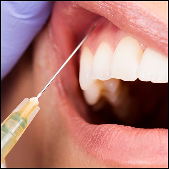
Eshealthtips.com – The retraction instrument is used to bring a needle to the site of injection from the contralateral premolar region. The operator holds the syringe at a 45-degree angle to the long axis of the tooth and bevels the needle to face the apex of the tooth root. The needle is then advanced a short distance until it makes contact with the bone. The needle is then pulled back by a few millimeters and the injection is completed.
Treatments Done on Teeth
In half of the cases, the foramen is located in the posterior third molar. In the other half, it is positioned medial to the second molar. In either case, the foramen is 1-2 mm anterior to the third molar. After identifying the injection site, the cotton swab should be positioned on the tissue until it blanches. Then, the syringe should be aimed perpendicular to the injection site.
Another site of injection is the nasopalatine (NP) area. In this technique, the dental practitioner injects a local anesthetic into the palatal premaxilla. The NP area is a region of soft tissue, enabling treatment to be performed in the innervated tooth. This injection site is commonly called local infiltration. The NP site can be visualized through the posterior aspect of the incisor.
In order to ensure a comfortable and safe injection experience, the dentist should raise the pH of the local anesthetic solution in the dental cartridge to a pH of 7.4. This will prevent a ‘burning’ sensation caused by the anesthetic agent. It also increases the number of B molecules available to reach the nerve. The more B molecules that reach the nerve, the deeper the anesthetic effect.
Procedures Involving Injecting Anesthesia
Another option for numbing a tooth is to inject a local anesthetic agent through the gums. This is called cutaneous infiltration. This procedure involves injecting the anesthetic directly into the site. Nerve blocks and field blocks are other options. Nerve blocks target specific areas of the body, and can be easily spotted by a landmark. In this technique, the dentist can inject an anesthetic directly into a single site or multiple locations.
The location of the intraoral needle is the most common site for numbing. This site is 2 mm apical to the gingival margin of the teeth. It should not be located near the occlusal plane, and should not touch the maxillary bone. The needle is then inserted into the soft tissue until it reaches the bone. The depth of penetration varies from individual to individual. The anesthetic solution is deposited in a few millimeters of bone.
The intraosseous technique is performed with a special tool. It is known for its fast onset and minimal side effects. In addition, this technique does not numb the lips and tongue and is considered an adjunctive method after nerve block. Contraindications include severe inflammation or infection in the area. It is a relatively safe method of local anesthesia. Its onset is similar to the standard mandibular block.
Most Common Anesthesia Injection Technique
In the most common anesthetic injection technique, the IAN, or inferior alveolar nerve, is anesthetized. This technique affects all the teeth on one side of the mandible, as well as the bone from the inferior ramus to the midline. This technique can also affect the lingual soft tissue and periosteum of the mandible, as well as the anterior two-thirds of the tongue and the floor of the oral cavity.
In the intraosseous method, the needle is advanced to a length of one-half to three-fourths of its length, where it makes contact with the molars. A local anesthetic solution is then deposited in the area over a period of one minute, anesthetizing the long buccal and inferior alveolar nerves. The ipsilateral mandibular teeth, along with associated hard and soft tissue, are also anesthetized with local anesthetic. The floor of the mouth and the anterior two-thirds of the tongue are also anesthetized with local anesthetic.

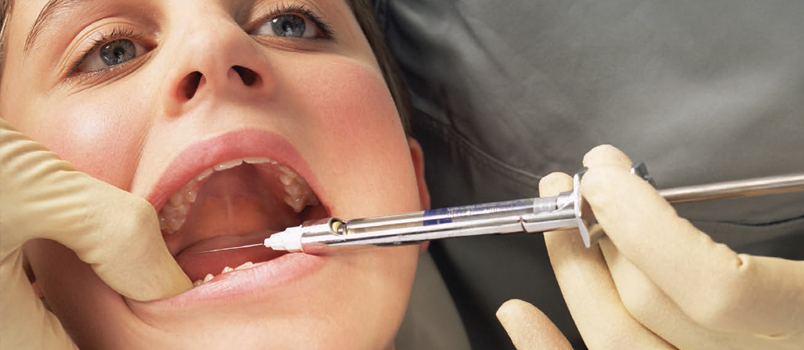
The injection is performed by opening the mouth maximally. The lingual portion of the maxillary second molar is a reference point for the height of the injection. The needle is then moved distally, parallel to the imaginary line drawn from the intertragic notch to the corner of the mouth. The needle is removed after the injection is completed. The procedure is complete with minimal pain. Once the injection has been completed, the patient will be fully sedated.