Eshealthtips.com – The bones of the wrist make up most of the skeletal structure of the hand. These bones allow different tendons and neurovascular structures to reach the wrist. Moreover, the bones also provide innervation and blood supply. Let’s learn more about these bones. Let’s begin with a brief description. The bones of the wrist provide support for the tendons and muscles attached to them. They also serve as important parts of the hand.
The Eight Bones That Make Up the Human Wrist
There are eight bones that make up the human wrist. Each is connected to the other through ligaments. Each finger has two phalanges and a carpal bone. In the coronal plane, these bones form an arch. The flexor retinaculum connects the lateral and medial sides of the arch. The hamate also serves as a joint for different muscles. Therefore, if the hamate is fractured, the blood flow can be disrupted.
The human wrist is made up of eight small bones. The ulna is found on the side of the forearm with a small finger. Unlike the radius, the ulna does not twist. Therefore, the ulna remains in the same position whenever the hand changes positions. The ulna has joint structures at the wrist and elbow. The ulna and humerus joint is a hinge-type joint. However, the smaller surface area of the ulna makes it more vulnerable to breakage.
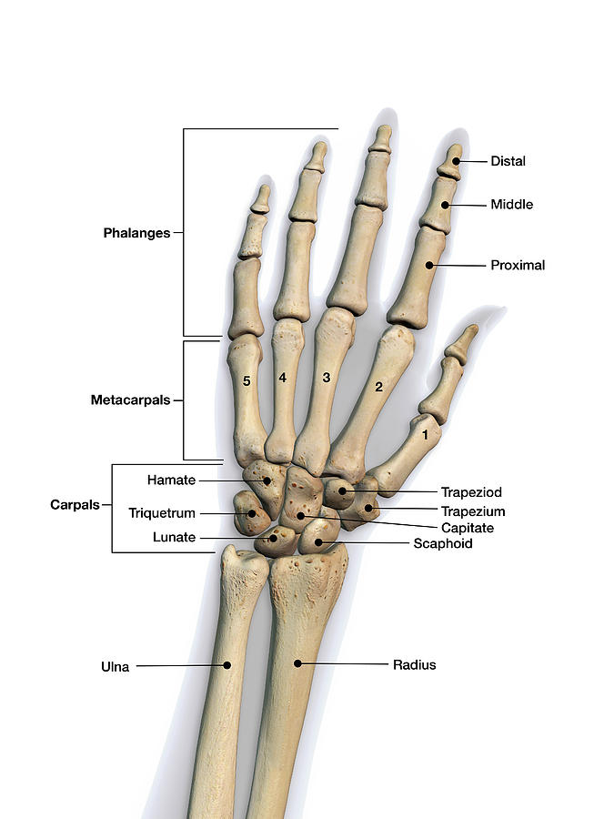
Generally, the bones of the hand are grouped into three categories. These groups are called carpus. The carpus consists of eight bones, which form the wrist and the root of the hand. The digits are made up of phalangeal bones. These bones are arranged in two rows, one on the proximal side and one on the distal side. The carpal bones of the more proximal row articulate with the ulna and radius. The intercarpal joints are joined by ligamentous attachments.
The Capitate is the Big Bone in the Wrist
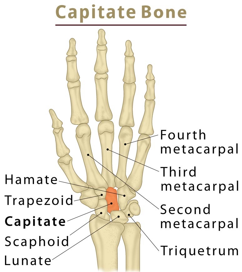
The capitate is the large bone in the wrist. It forms the joints with several other bones in the hand and wrist. The capitate sits beneath the middle finger metacarpal bone and contributes to the wrist motion. The hamate also articulates with the capitate in a functional way. It is important to have intact capitates and haematoid if you want to function properly.
Unlike other fossilized remains, modern humans and Neandertals shared the same carpal complex. The modern human carpal complex involves palmar broadening and repositioning of the trapezoid, as well as novel enlargement of the trapezoid-capitate articulation. Moreover, trapezium facets engulf the distoradial aspect of the scaphoid tubercle, which is the most modern human wrist.
A new study has revealed the relationship between bone density and vibration frequency. The results of the study are based on the imaging of a wrist bone taken at 41 keV. The rescaled rocking curves are flatter for dense trabecular struts. The trabeculae are responsible for a greater amount of reflected X-rays at larger angles. These results indicate that these devices could help doctors distinguish between fracture and strain.
The Fingers and Ulna are the Most Common Bones in the Wrist
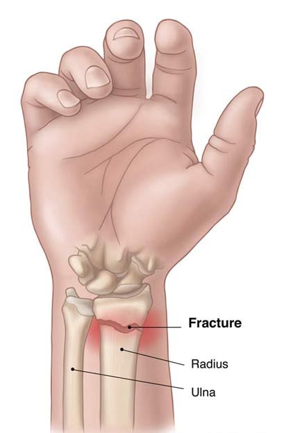
The wrist is made up of eight bones, including the radius, the ulna, and the carpals. These bones are connected to two long bones in the arm. The radius and the ulna are the most common bones in the wrist. The ulna is shorter and thinner, and the radius is the thickest. These two bones connect to each other on the thumb and pinky sides of the wrist.
Three major nerves supply the muscles of the hand. Table 3 shows their distribution. Each major nerve trunk has many branches, which diverge into numerous smaller ones. As a result, the neuromuscular system is coordinated in the brain, where the response to stimulus is subconscious and reflex. For example, if an object is slipping, we will automatically grip it more tightly and reject it. The same goes for noxious stimuli.
Reference: