Eshealthtips.com – The radial nerve is the largest nerve of the upper limb, which arises from the posterior cord of the brachial plexus in the axillary region. It travels down the outside of the forearm, supplying sensation to the radial 3 and 1/2 digits and the hand. The radial nerve has two branches, superficial and deep. The superficial branch supplies sensory information to the skin of the forearm and the deep branch stimulates muscles to move upward.
Experiencing Pain and Weakness in Left Shoulder
The senior author treated a patient who presented with pain and weakness in the left shoulder and radial nerve. She showed a 5/5 strength level in all muscles, 0/5 strength in the triceps, and numbness over the first dorsal web space in the radial nerve distribution. Her brachial plexus MRI and EMG were normal. However, EMG revealed a fibrillation pattern of the radial nerve muscles.

The ulnar artery and radial nerve are located in the cubital fossa, a hollow in the elbow. The radial nerve is made up of the ulnar nerve and musculocutaneous nerve, which run through the biceps brachii tendon and triceps brachii, as well as several tendons. These tendons, along with the biceps brachii, are responsible for the movements of the elbow, forearm, and shoulder.
FCR Incision Between Radial and Brachioradialis Nerves
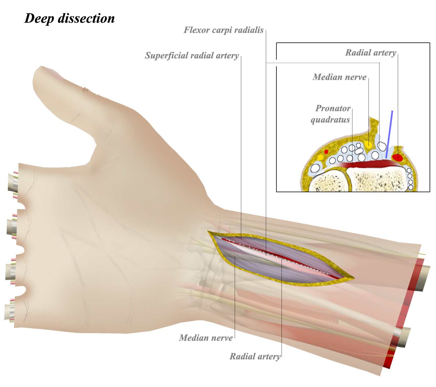
The anterior forearm approach exploits the intraosseous plane between the brachioradialis and flexor carpi radialis. Pronator is palpable along the radial forearm. The FCR is incised between the radial nerve and brachioradialis, while the abductor pollicis and extensor digitorum communis are mobilized laterally.
The interosseous membrane runs from the medial border of the radius to the lateral border of the ulna. It extends about one cm distal from the DRUJ, and its fibers are oriented at 60-degree angles. They are most taut in neutral to 30deg supination and relaxed in pronation. They act as a landmark in the radial compartment, providing a surgical reference for positioning the ulna.

The median nerve is attached to the humerus and is in relation to the medial side of the artery for 2.5 cm. The radial nerve runs parallel to the medial side of the humerus, and it innervates upper limb muscles. The radial nerve was the least likely to be injured at this location and the tendons that attach to the humerus are intrinsic to the hand.
The Most Prominent and Deepest Muscles of the Forearm
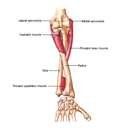
The supinator muscle is the most prominent and deepest muscle of the forearm. Supination of the forearm moves the PIN away from the incision, protecting it. The supinator muscle is raised subperiosteally and medially, and moves posteriorly around the radial neck. The supinator is usually attached to the PIN, which is a danger if the supinator is cut in the anterior approach.
The forearm muscles may also contribute to isolated radial nerve palsy. This may be due to a variant anatomy that creates more tension on the nerve during dislocation. It is important to preserve the cuff of tissue so that muscle repairs can occur. The radial nerve itself is often protected in such a way. And a shoulder dislocation may also damage the radial nerve. However, there is no definite evidence that the forearm muscles are involved in all cases of radial nerve palsy.

An anterior approach to the radius is usually ineffective, owing to its subcutaneous surface. The ulna, on the other hand, can be exposed entirely along its subcutaneous border. Surgical approaches that involve posterior and anterior approaches to the radius do not provide adequate exposure. Moreover, the inner nervous plane of the ulna makes it possible to expose the entire ulna with a single incision.
Reference: