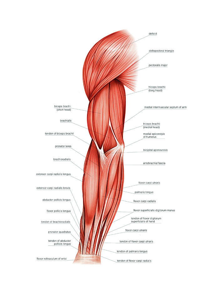
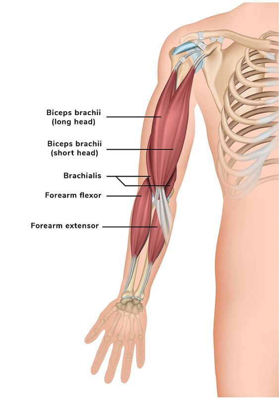
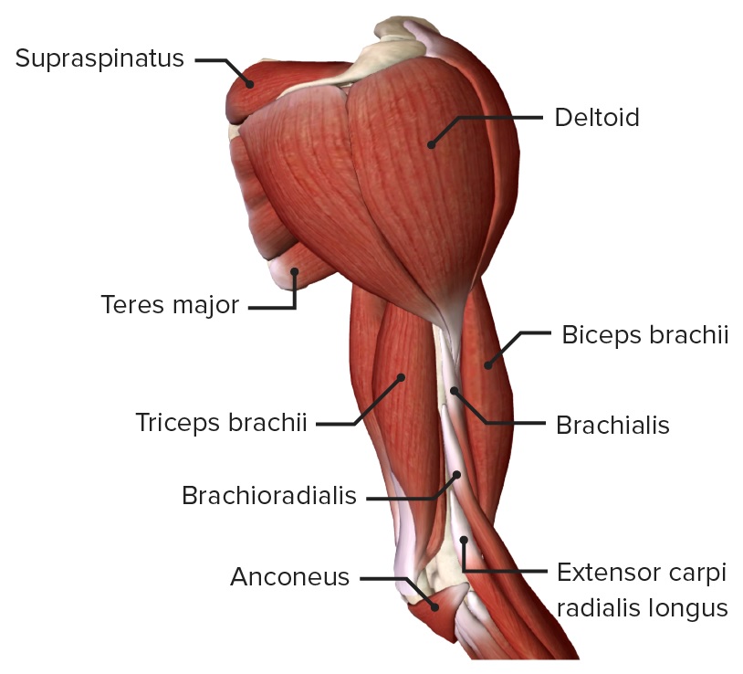
Eshealthtips.com – The muscles of the Upper Arm are made up of three major groups: the biceps, triceps, and brachialis. There are several nerves that run through these muscles, which can cause pain in the upper arm. Pain in the UpperArm is a symptom of a variety of conditions, including neck, shoulder, and rotator cuff injuries. Tendonitis can occur at the insertion point of the biceps or triceps into the elbow.
Anatomy of the Upper Arm Consists of Four Major Muscles
The anatomy of the UpperArm is made up of four major muscles: the biceps brachii, triceps brachii, and biceps femoris. The biceps brachii is located in the anterior compartment and is innervated by the musculocutaneous nerve. The triceps femoris and triceps teres are located in the posterior compartment.
The anatomy of the UpperArm is very complicated. The four muscles located in the UpperArm are grouped into two compartments: the anterior and the posterior. The anterior compartment is supplied by the brachial artery and contains the biceps and the triceps brachii, the biceps femoris, and the triceps digitorum profundus. The arm is made up of the extensor carpi radialis and the flexor digitorum longus.

The upper arm is divided into three compartments: the anterior compartment is in front of the humerus, while the posterior compartment is behind the humerus, the main bone in the upper arm. The biceps brachii, commonly referred to as the biceps, is a muscle with two heads in the shoulder region. This muscle helps with the flexion of the upper arm. The triceps brachii is another muscle located in the anterior area.
Various Upper Muscle Functions
The muscles of the Upper Arm are important for a variety of functions. For example, they control the top joints of the fingers, turn the palm of the hand toward the body, raise the arm above the head, and rotate the forearm. The three tendons are located in the anterior and posterior compartments of the upper arm. The radialis brachii is the biceps muscle. The triceps brachii is the second. The biceps muscles are located in the posterior compartment.
The upper arm contains four muscles: the biceps brachii, the triceps brachii, and the biceps. The biceps brachii, also called biceps, is the largest of the four and is the most prominent muscle in the Upper Arm. Its two heads near the shoulder are responsible for the flexion of the upper arm. The triceps and adductor tendons in the shoulder help to stabilize the humerus and glengeal bones.

There are two types of tendons in the upper arm: the biceps brachii and the teres minor. The latter is the thicker and longer version. Its actions and innervation are different in the two muscles. The biceps brachii is the lateralmost muscle. The triceps is the smallest of the three. Both are located in the posterior compartment. While the triceps brachii is the largest of the three, it is also the only one to be in the posterior compartment.
Triceps and Brachialis are Responsible for Upper Arm Flexion
There are two types of tendons in the Upper Arm. The biceps brachii is the more common type and has a musculocutaneous nerve. The biceps brachioiis is the larger of the two types, but it can also be the most complex. This artery is responsible for all movements of the upper arm. It is connected to the shoulders, which in turn connects the muscles.

The muscles in the Upper Arm are responsible for a variety of movements in the body. Generally, the upper arm is divided into two compartments: the anterior compartment is located near the shoulder and the posterior compartment is behind the elbow. In both compartments, there are muscles that act on the humerus, a main bone in the UpperArm. The biceps brachii is also called the biceps and is a rotator cuff muscle.
Reference: